Overview
- Peptide (C)NGVPESTSTDTPPDIDLHN, corresponding to amino acid residues 392-410 of human Kir2.1 (Accession P48049). Intracellular, C-terminal part.
- Rat brain and rat heart membranes (1:200). Addition of 0.1% Tween to the standard milk block is recommended with this lot.
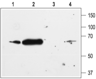 Western blot analysis of rat heart (lanes 1 and 3) and rat brain (lanes 2 and 4) membranes:1,2. Anti-Kir2.1/KCNJ2 Antibody (#APC-026), (1:200).
3,4. Anti-Kir2.1/KCNJ2 Antibody, preincubated with Kir2.1/KCNJ2 Blocking Peptide (#BLP-PC026).
- Transfected HEK 293 cell lysate (Preisig Muller, R. et al. (2002) Proc. Natl. Acad. Sci. U.S.A. 99, 7774.).
- Rat brain sections.
- Mouse ventricular myocytes (1:200) (Clark, R.B. et al. (2001) J. Physiol. 537, 979.).
- Human bladder urothelial cells (BUC) (Sun, Y. et al. (2007) Am. J. Physiol. 292, C106.).
Kir2.1 is a member of the family of inward rectifying K+ channels. The family includes 15 members that are structurally and functionally different from the voltage-dependent K+ channels.1
The family’s topology consists of two transmembrane domains that flank a single and highly conserved pore region with intracellular N- and C-termini. As is the case for the voltage-dependent K+ channels the functional unit for the Kir channels is composed of four subunits that can assemble as either homo- or heterotetramers.
Kir channels are characterized by a K+ efflux that is limited by depolarizing membrane potentials thus making them essential for controlling resting membrane potential and K+ homeostasis.
Kir2.1 is a member of the Kir2.x subfamily that includes four members (Kir2.1- Kir2.4) that are characterized by strong inward rectification and high constitutive activity.
Kir2.1 is expressed in a variety of tissues including the heart, brain, vascular smooth muscle cells and skeletal muscles.
In the heart, Kir2.1 is a molecular component of the IK1 current that is responsible for setting the resting membrane potential, preventing membrane hyperpolarization due to Na+ pump activity, influencing propagation velocity, altering the electrical space constant, and promoting late phase repolarization.2 In fact, mutations in Kir2.1 channels have been linked to a form of long QT syndrome (LQT7) known as Andersen's syndrome that is characterized by cardiac arrhythmias, periodic paralysis, and dysmorphic features.3
 Antibody")
Expression of Kir2.1 in canine myocytes.
Immunocytochemical staining of canine myocytes using Anti-Kir2.1/KCNJ2 Antibody (#APC-026). Kir2.1 staining (green) is detected in both ventricle (V) and atria (A). Kir2.1 staining is eradicated when antibody is incubated with the peptide antigen (C and F).Adapted from Melnyk, P. et al. (2002) Am. J. Physiol. 283, 1123. with permission of The American Physiological Society.