Overview
- Peptide (C)THHFKDAFSGRDTS, corresponding to amino acid residues 218-231 of mouse xCT/SLC7A11 (Accession Q9WTR6). 3rd extracellular loop.
- Mouse J774 macrophage cells; human THP-1 monocytic leukemia cells (2.5 µg).
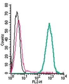 Cell surface detection of xCT/SLC7A11 in live intact mouse J774 macrophage cells:___ Cells.
___ Cells + Rabbit IgG isotype control-PE.
___ Cells + Anti-xCT/SLC7A11 (extracellular)-PE Antibody (#ANT-111-PE) (2.5 µg).
The SLC7 family is divided into two subgroups, the cationic amino acid transporters (the CAT family, SLC7A1–4) and the glycoprotein-associated amino acid transporters (the gpaAT family, SLC7A5–11) also called light chains or catalytic chains of the hetero(di)meric amino acid transporters (HAT). The gene SLC7A11 encodes the xCT protein (also known as Cystine/glutamate transporter).
xCT, as well as other HATs, is comprised of a light chain with 12 non-glycosylated transmembrane helices and is associated with the glycosylated heavy chain 4F2hc (CD98, SLC3A2) or rBAT (SLC3A1) through a conserved disulphide bridge. The COOH-terminus of the xCT is localized intracellularly. Association of the light and heavy chains is required for surface expression of the transporter which forms the heterodimeric amino acid transport system xc−. xCT is mainly expressed macrophages, brain, retinal pigment cells, liver and kidney. xCT expression is elevated in cells requiring high glutathione synthesis, such as activated macrophages, neuronal and glial cells and other cells after glutathione depletion1.
xCT operates in a Na+-independent and electroneutral mode, exchanging extracellular anionic cystine (with pH dependence) for glutamate with a stoichiometry of 1:1. The driving force for this exchange is generated by the cystine concentration gradient (intracellular reduction) and the high intracellular concentration of glutamate.
xCT has been suggested as a mediator of treatment resistance in cancer patients. In tumor cells, xCT plays a crucial role in regulating intracellular levels of glutathione which has been broadly implicated in resistance to chemotherapy2.