Overview
Cav1.3/CACNA1D Blocking Peptide (#BLP-CC005) is the original antigen used for immunization during Anti-CaV1.3 (CACNA1D) Antibody (#ACC-005) generation. The blocking peptide binds and ‘blocks’ Anti-Cav1.3/CACNA1D primary antibody, this makes it a good negative reagent control to help confirm antibody specificity in western blot and immunohistochemistry applications. This control is also often called a pre-adsorption control.
Applications
 Western blot analysis of rat brain membranes:1. Anti-CaV1.3 (CACNA1D) Antibody (#ACC-005), (1:200).
2. Anti-CaV1.3 (CACNA1D) Antibody, preincubated with Cav1.3/CACNA1D Blocking Peptide (#BLP-CC005).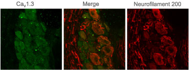 Expression of CaV1.3 in rat DRGImmunohistochemical staining of adult rat dorsal root ganglion (DRG) with Anti-CaV1.3 (CACNA1D) Antibody (#ACC-005). Staining appears in clusters of cells (green) but not in axons. Staining of neurofilament 200 demonstrates partial overlap with CaV1.3 in neuronal staining but not in axons (red).
 Western blot analysis of mouse brain (lanes 1 and 4), rat brain (lanes 2 and 5) and human brain neuroblastoma cells (SH-SY5Y) (lanes 3 and 6):
1-3. Guinea pig Anti-CaV1.3 (CACNA1D) Antibody (#ACC-005-GP), (1:200).
4-6. Guinea pig Anti-CaV1.3 (CACNA1D) Antibody, preincubated with Cav1.3/CACNA1D Blocking Peptide (#BLP-CC005).
Following a broad screen of secondary antibodies, the following were used for this application:
Western blot analysis:
#106-035-006 (Jackson ImmunoResearch)
#A7289 (Sigma) Expression of CaV1.3 in rat DRGsImmunohistochemical staining of rat dorsal root ganglion (DRG) using Guinea pig Anti-CaV1.3 (CACNA1D) Antibody (#ACC-005-GP), (1:200). A. CaV1.3 staining (red) is detected in some DRG cells (arrows). B. Nuclei staining using DAPI as the counterstain. C. Merged image of panels A and B.
Properties
- (C)DNKVTIDDYQEEAEDKD, corresponding to amino acid residues 859-875 of rat CaV1.3 (Accession P27732).