Overview
- Peptide (C)RNLRQIFQSLPPFMD, corresponding to amino acid residues 221-235 of rat TPCN1 (Accession Q9WTN5). 2nd luminal loop.
- Rat heart lysate and liver membranes (1:200). Addition of 0.1%-0.5% Tween-20 to antibody solution is highly recommended.
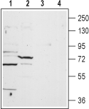 Western blot analysis of rat heart lysate (lanes 1 and 3) and liver membrane (lanes 2 and 4):1,2. Anti-TPCN1 Antibody (#ACC-071), (1:200).
3,4. Anti-TPCN1 Antibody, preincubated with TPCN1 Blocking Peptide (#BLP-CC071).
- Rat lung paraffin embedded sections (1:100).
Among various vertebrate species, three genes are known to encode two-pore segment channels (TPCs) termed TPC1-3. Interestingly TPC3 seems to be absent from the genomes of primates and rodents1. The primary sequence of these channels indicates the presence of two putative pore-forming repeats. Each repeat contains six transmembrane domains and a pore loop, a structure strikingly reminiscent of many voltage-gated Na+ (Nav) and Ca2+ (Cav) channels2. These twelve transmembrane structures are further thought to form functional dimers3. Both TPC1 and TPC2 show ubiquitous expression, while that of TPC1 is exceptionally high in spleen, lung, liver, and kidney2.
Ca2+-mobilizing messengers such as inositol triphosphate, cyclic ADP ribose and nicotinic acid adenosine dinucleotide phosphate (NAADP) are responsible for the intracellular changes in Ca2+ ion concentration4.
In contrast to the other Ca2+-mobilizing agents, NAADP, the most potent of these Ca2+ releasing molecules increases the cytosolic Ca2+ concentration via Ca2+ channels located on acidic vesicles (endolysosomes)5,6. Only quite recently, after almost a decade of being cloned, TPC1 and TPC2 were both found to be responsible for the NAADP-induced release of Ca2+ 7,8. Evidence that these two channels are indeed responsible for the release of Ca2+ is quite compelling since overexpression of TPC1 and its knockdown or point mutation of a critical residue increase and exacerbate Ca2+ release respectively7. In addition, b-cells from TPC2 knockout mice exhibited no Ca2+ release from endolysosomes upon NAADP stimulation8. Finally, in a study using immunopurified channels, it was demonstrated that TPC1 and TPC2 both respond to very low concentrations of NAADP and are unequivocally responsible for the release of Ca2+, whereas TPC3 may negatively regulate the release of Ca2+ 9.
As these channels have only recently been discovered, very little is known about their physiology and gating mechanisms. Their probable involvement in a number of diseases such as lysosomal storage disease (LSDs), caused by the dysfunction of lysosomal associated proteins, has yet to be deciphered1.