Overview
Type: Synthetic peptide
Form: Lyophilized powder
GFAP Blocking Peptide (#BLP-FP001) is the original antigen used for immunization during Anti-GFAP Antibody (#AFP-001) generation. The blocking peptide binds and ‘blocks’ Anti-GFAP primary antibody, this makes it a good negative reagent control to help confirm antibody specificity in western blot and immunohistochemistry applications. This control is also often called a pre-adsorption control.
Applications: wb, ihc
For research purposes only, not for human use
Applications
Demonstration of Pre-adsorption control
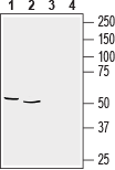 Western blot analysis of rat brain membranes (lanes 1 and 3), (1:2000) and mouse brain lysate (lanes 2 and 4), (1:400):1,2. Anti-GFAP Antibody (#AFP-001).
3,4. Anti-GFAP Antibody, preincubated with GFAP Blocking Peptide (#BLP-FP001).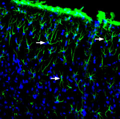 Expression of GFAP in rat brainImmunohistochemical staining of free-floating rat frozen brain sections using Anti-GFAP Antibody (#AFP-001), (1:2000). In rat parietal cortex, GFAP immunoreactivity (green) appears in astrocytes (arrows). Nuclei are stained using DAPI as the counterstain (blue).
 Multiplex staining of GFAP and AQP4 in rat brainImmunohistochemical staining of immersion-fixed, free floating rat brain frozen sections using Anti-GFAP Antibody (#AFP-001), (1:2000) and Anti-Aquaporin 4 (AQP4) (300-314)-ATTO Fluor-594 Antibody (#AQP-014-AR), (1:100). A. In rat fornix, GFAP immunoreactivity (green) appears in astrocytic processes. B. In same section, AQP4 staining (red) is detected in blood vessels. C. Merge of panel A and panel B shows colocalization around a large blood vessel (arrows). Nuclei are stained using DAPI as the counterstain (blue).
 Multiplex staining of Connexin-43 and GFAP in rat cerebellum.Immunohistochemical staining of perfusion-fixed frozen rat brain sections with Guinea pig Anti-Connexin-43 Antibody (#ACC-201-GP), (1:200), followed by goat anti-guinea pig-AlexaFluor-594 and Anti-GFAP Antibody (#AFP-001), (1:1200), followed by goat anti-rabbit-AlexaFluor-488. A. Connexin-43 immunoreactivity (red) appears as positive puncta (arrows). B. GFAP immunoreactivity (green) appears along the Bergmann glial processes (arrows point at examples). C. Merge of the two images shows CNX-43 puncta distributed along the Bergmann glial processes. Cell nuclei are stained with DAPI (blue).
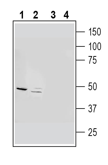 Western blot analysis of rat brain synaptosome lysates (lanes 1 and 3) and mouse brain lysates (lanes 2 and 4):1-2. Guinea Pig Anti-GFAP Antibody (#AFP-001-GP), (1:500).
3-4. Guinea Pig Anti-GFAP Antibody, preincubated with GFAP Blocking Peptide (BLP-FP001).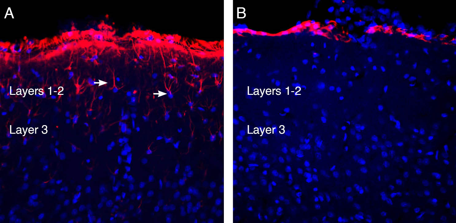 Expression of GFAP in rat parietal cortex.Immunohistochemical staining of perfusion-fixed frozen rat brain sections with Guinea Pig Anti-GFAP Antibody (AFP-001-GP). Sections were incubated with guinea pig anti GFAP (#AFP-001-GP), (1:1200), followed by goat anti guinea pig conjugated with Alexa 594 (red). A, GFAP immunoreactivity appears in cortical astrocytes in layers 1-2 (arrows). B, Pre-incubation of the antibody with GFAP blocking peptide (BLP-FP001), suppressed staining. Cell nuclei are stained with DAPI (blue).
 Multiplex staining of CLEC7A and GFAP in rat hippocampal CA1 regionImmunohistochemical staining of perfusion-fixed frozen rat brain sections with Anti-CLEC7A/Dectin-1 (extracellular) Antibody (#ALR-062), (1:600), followed by goat anti-rabbit-AlexaFluor-488 and Guinea Pig Anti-GFAP Antibody (#AFP-001-GP), (1:600), followed by goat anti-guinea pig-AlexaFluor-594. A. CLEC7A immunoreactivity (green) appears in astrocyte profiles. B. GFAP immunoreactivity (red) appears in astrocyte profiles. C. Merge of the two images demonstrates extensive co-localization. Cell nuclei are stained with DAPI (blue). P = pyramidal layer.
Properties
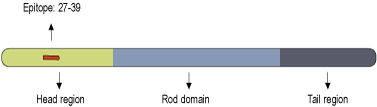
Sequence
- (C)RHLGTIPRLSLSR, corresponding to amino acid residues 27-39 of rat Glial fibrillary acidic protein (Accession P47819).
Accession (Uniprot) Number P47819
Peptide Confirmation Confirmed by amino acid analysis and mass spectrometry.
Purity >70%
Storage Before Reconstitution Lyophilized powder can be stored intact at room temperature for two weeks. For longer periods, it should be stored at -20°C.
Reconstitution 100 µl double distilled water (DDW).
Concentration After Reconstitution 0.4 mg/ml.
Storage After Reconstitution -20°C.
Antigen Preadsorption Control 1 µg peptide per 1 µg antibody.
Standard Quality Control Of Each Lot Western blot analysis.