Overview
Type: Synthetic peptide
Form: Lyophilized powder
Synaptophysin Blocking Peptide (#BLP-NR013) is the original antigen used for immunization during Anti-Synaptophysin Antibody (#ANR-013) generation. The blocking peptide binds and ‘blocks’ Anti-Synaptophysin primary antibody, this makes it a good negative reagent control to help confirm antibody specificity in western blot and immunohistochemistry applications. This control is also often called a pre-adsorption control.
Applications: wb, ihc
For research purposes only, not for human use
Applications
Demonstration of Pre-adsorption control
 Western blot analysis of rat brain membranes (lanes 1 and 4), mouse brain membranes (lanes 2 and 5) and rat PC12 pheochromocytoma cell line lysate (lanes 3 and 6):1-3. Anti-Synaptophysin Antibody (#ANR-013), (1:400).
4-6. Anti-Synaptophysin Antibody, preincubated with Synaptophysin Blocking Peptide (#BLP-NR013).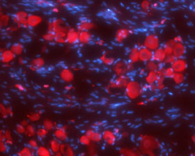 Expression of Synaptophysin in rat DRGImmunohistochemical staining of rat dorsal root ganglia (DRG) frozen sections using Anti-Synaptophysin Antibody (#ANR-013), (1:100). SYP (red) is expressed in DRG neurons. Hoechst 33342 (blue) shows nuclear staining and is used as the counterstain.
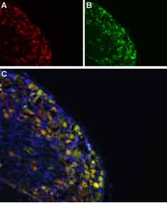 Colocalization of NaV1.8 and Synaptophysin in rat DRGImmunohistochemical staining of rat DRG frozen section using Anti-NaV1.8 (SCN10A)-ATTO Fluor-594 Antibody (#ASC-016-AR) and Anti-Synaptophysin Antibody (#ANR-013). A. NaV1.8 staining (red). B. SYP staining (green). C. Merged image demonstrates a partial overlap in the distribution of NaV1.8 and SYP within the DRGs. DAPI is used as the counterstain (blue).
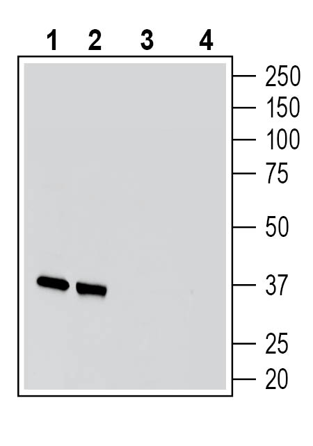 Western blot analysis of rat brain membranes (lanes 1 and 3) and mouse brain membranes (lanes 2 and 4):1-2. Guinea Pig Anti-Synaptophysin Antibody (#ANR-013-GP), (1:200).
3-4. Guinea Pig Anti-Synaptophysin Antibody, preincubated with Synaptophysin Blocking Peptide (BLP-NR013).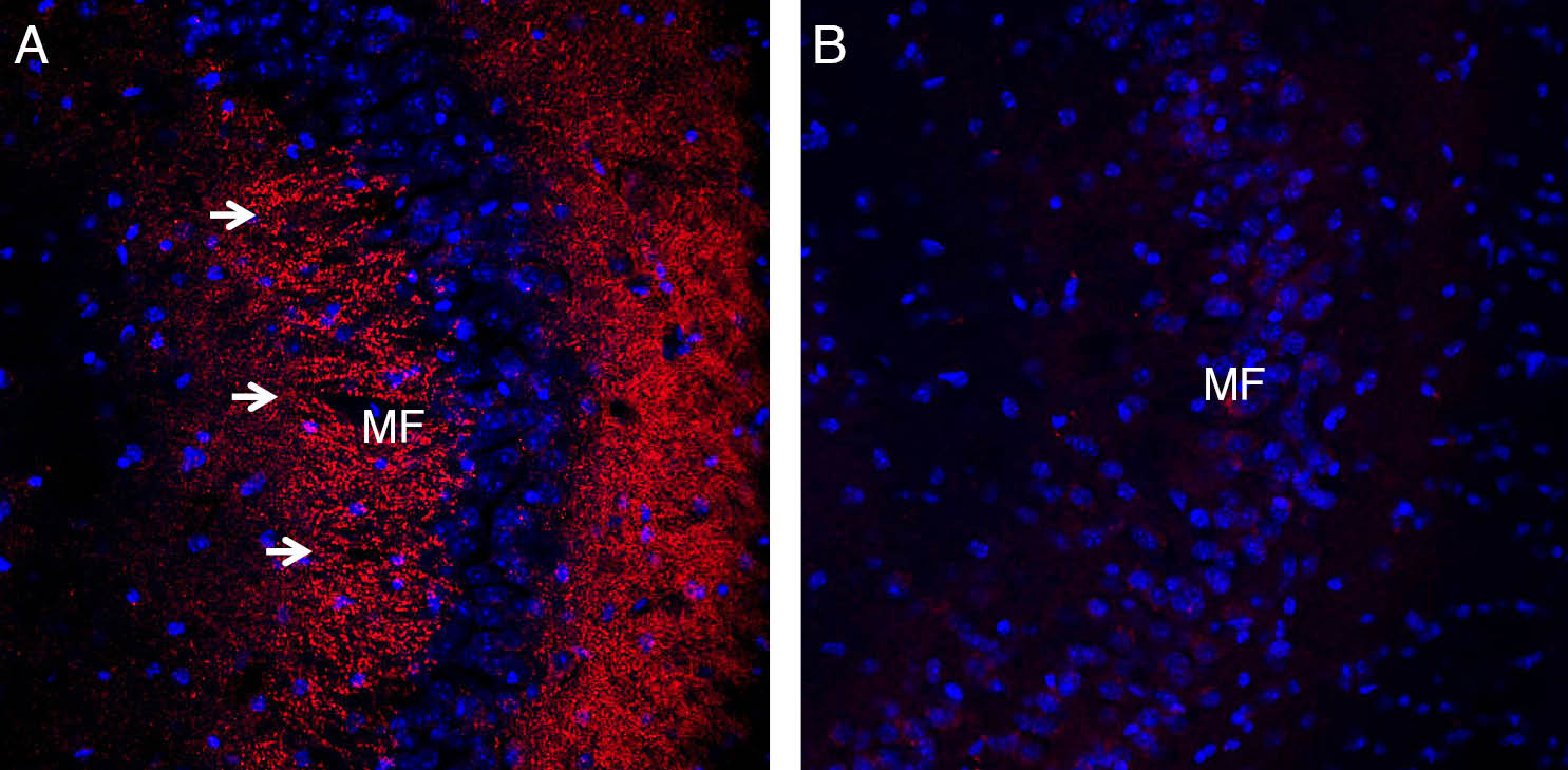 Expression of Synaptophysin in mouse hippocampus.Immunohistochemical staining of perfusion-fixed frozen mouse brain sections with Guinea Pig Anti-Synaptophysin Antibody (#ANR-013-GP), (1:200), followed by goat anti-guinea pig-AlexaFluor-594. A. Staining in the hippocampal CA3 region, showed immunoreactivity (red) in the mossy fiber terminal zone (MF, arrows). B. Pre-incubation of the antibody with Synaptophysin Blocking Peptide (BLP-NR013), suppressed staining. Cell nuclei are stained with DAPI (blue).
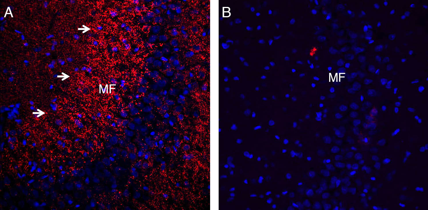 Expression of Synaptophysin in rat hippocampus.Immunohistochemical staining of perfusion-fixed frozen rat brain sections with Guinea Pig Anti-Synaptophysin Antibody (#ANR-013-GP), (1:200), followed by goat anti-guinea pig-AlexaFluor-594. A. Staining in the hippocampal CA3 region, showed immunoreactivity (red) in the mossy fiber terminal zone (MF, arrows). B. Pre-incubation of the antibody with Synaptophysin Blocking Peptide (BLP-NR013), suppressed staining. Cell nuclei are stained with DAPI (blue).
Properties
Sequence
- CRQTGNT(S)KELRD, corresponding to amino acid residues 178-190 of rat synaptophysin (Accession P07825).
Accession (Uniprot) Number P07825
Peptide Confirmation Confirmed by amino acid analysis and mass spectrometry.
Purity >70%
Storage Before Reconstitution Lyophilized powder can be stored intact at room temperature for two weeks. For longer periods, it should be stored at -20°C.
Reconstitution 100 µl double distilled water (DDW).
Concentration After Reconstitution 0.4 mg/ml.
Storage After Reconstitution -20°C.
Antigen Preadsorption Control 1 µg peptide per 1 µg antibody.
Standard Quality Control Of Each Lot Western blot analysis.